Google Ad space
finances and sponsors ENT USAsm Website. ENT USAsm,
Cumberland Otolaryngology or Dr Kevin Kavanagh, MD do not endorse,
recommend, referrer to or are responsible for the Advertisements or
for the
content or claims made
in the Advertisements.
For nasal and nose allergies,
tonsil, adenoid and ear tube surgery,
ear infections and videos:
Go
To www.entusa.com
For Continuing Medical Education Credits: Go
To www.cme-usa.org
External Ear: Contains the external auditory canal and
auricle (outer ear).
Middle Ear and Eardrum (tympanic membrane) and three ear bones or
ossicles: malleus, incus and stapes.
Inner Ear: Contains the cochlea, a snail-shaped bone which
transforms sounds into nerve impulses, the vestibule, contains the utricle
and saccule which sense motion in relation to gravity and three semicircular
canals which sense rotational motion.
Mastoid Air Cells: These are air cells which are behind the
ear canal and middle ear.
The web pages contained in this section present
60 temporal bone anatomy slides. Histology
sections from both the right and left temporal bone are
available. There are 10 histological sections per page.
The first page starts in the mid-temporal bone at the level of the
stapes. Each histology section can be viewed in three sizes:
thumbnail, 750 pixels wide and 3000 pixels wide. The 3000 pixels
format is approximately 400 Kbytes in size and may take 3 to 4 minutes to view
using a 28 baud modem. To view the 700 pixel format, click on the
thumbnail, to view the 3000 pixel version click on the the "View Highest
Resolution" option next to the thumbnail. View
Right Temporal Bone SlidesView
Left Temporal Bone Slides
The three middle
ear bones are called Ossicles. They are the Malleus (attaches to the Ear
Drum), Incus (transmits sound between the Malleus and Stapes), and
stapes which transmits sound to the Vestibule of the inner ear.
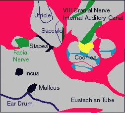
Slide of Stapes Footplate,
Vestibule (Utricle and Saccule), Cochlea, dehiscent Facial (VII) Nerve,
Internal Auditory Canal, Malleus, Incus and Ear Drum.
The cochlea is composed of the Modiolus (Yellow) which transmits the
Auditory Nerve (VIII Cranial Nerve) to the Organ of Corti. The
Spiral Lamina and Ligament is shown in blue. (Macula of Saccule is
shown in dark green)
CLICK
ON ANATOMY SLIDE TO ENLARGE
The Cochlea is the organ of
hearing. It is snail shaped and has nearly 2.75 turns. A
close up view is provided in this slide. The Organ of Corti is
shown in green, the Stria Vascularis which is thought to produce Endolymph
is shown in brown, and the Vestibular (Reissner) Membrane is shown in
black. CD: Cochlear Duct which contains Endolymph.
The sense organs of the semicircular canals are called
Crista Ampullaris. They sense angular or rotational motion.
The Cupula extends from the tip of the Crista forming a diaphragm which
is thought to be reflected with movement of the Endolymph. Shown
here is a Crista and its Ampullary groove, transmitting the Ampullary
branch of the Vestibular Division of the Vestibulocochlear, VIII, Nerve.
The Macula Acustica Utriculi and Macula Sacculi are
the sense organs for gravity. These sense organs have Otoconia
which are little "rocks" that move in relationship to
gravity. These rocks can be dislodged by trauma and can enter the
Posterior Semicircular Canal giving rise to
positional vertigo.
Also note the Stapedial Footplate and dehiscent Facial, VII, Nerve.
The Right Temporal Bone has better histopathology with Basilar Membrane,
Vestibular (Reissners) Membrane, Spiral Ligament, Stria Vascularis, Utricule and Saccule seen
in most specimens. View Right Temporal
Bone Slides
The Left Temporal Bone has better orientation with the gross anatomy better
demonstrated. View Left Temporal
Bone Slides
The Basilar Membrane, Utricle and Saccule are seen in most specimens.
In the performance of a Stapedectomy, the stapes
footplate is fixed and must be removed. The relationship between the
footplate and inner ear structures is very important. The distance between
the stapedial footplate and the saccule varies from 1.0 to 1.4mm and from the
footplate to the utricle varies from 0.7 to 1.4 mm. In 25 specimens the
shortest distance between the footplate and the saccule was 0.8 mm and between
the footplate and the utricle was 0.38 mm.* In patients with Meniere's
disease the endolymph is under high pressure and the membranes of the utricle
and saccule are distended. Sometimes they reach the footplate.
Stapedectomy in these patients is very hazardous and there is a high risk of
postoperative deafness.
* Anson B.J. and Donaldson, J.A. Surgical Anatomy of The Temporal Bone and Ear.
W.B. Saunders Company p 282, 1973
For nasal and nose allergies, tonsil, adenoid and
ear tube surgery, ear infections and videos.
Go To:
www.entusa.com
For Tobacco Facts and Info on Smoking, Larynx Cancer and Quitting.
Go To:
www.tobacco-facts.info


